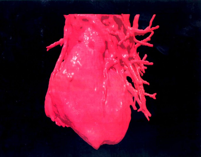
Three Dimensional (3 D Image Displays A Computerised Visualization Of A Human Heart (Photo : Getty Images)
Although American researchers were recently able to grow functional heart tissues from stem cells, growing hearts for transplants is still out of the question for now because of the lack of a skeletal frame to grow the heart.
However, 3D printed technology could copy the human heart and be used to guide surgeons in making fine cuts and sutures, especially when the patient is an infant and still has a small cardiac organ. That was what happened on March 11 in Jilin Province when surgeons at the People’s Hospital of Jilin operated on a nine-month-old baby.
Global Times reported that the baby was suffering from a severe congenital heart defect (CHD) which required an open heart surgery. The CHD involved a total pulmonary venous anomalous drainage in which all the four pulmonary veins of the heart were malpositioned. The infant also had atrial septal defect which caused blood to flow between the heart’s upper chambers.
As a result of these defects, the infant suffered from shortness of breath and weighed only 5.6kg before the procedure. Because the defect was rare and complicated and the patient so young and small, the doctors found it difficult to rely only on an ultrasound examination to develop the best surgery plan.
Zhang Xueqin, the hospital’s director of pediatric cardiac surgery center and the baby’s surgeon, and his team created, using 3D printing, a full-sized heart replica of the patient’s cardiac structure which helped them plan the surgery.
“With the model, we were able to know precisely where and how we should cut, and how big the incision should be. And with such a thorough plan, we spent only half the time we had expected to complete the surgery,” Zhang said.
The baby is recovering and has been transferred to the general ward. He is Jilin’s first cardiac surgery that used 3D printed technology, but the first in China was in July 21, 2015, also on a nine-month-old baby with CHD in Jiangsu Province.
To create 3D prints, surgeons used the Mimics software which imports MRI images and processes it to reconstruct a 3D rendered model that could communicate with a 3D printer. It usually takes a few hours to print a heart model, according to Biomedical.materialise.com.