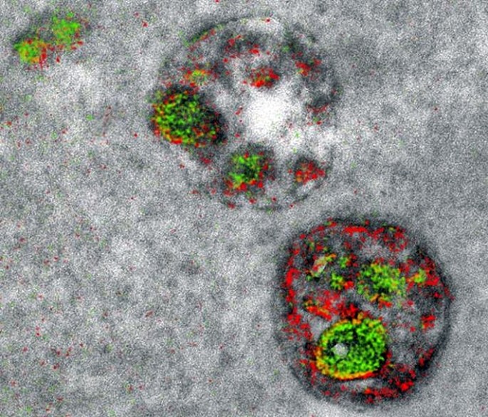
A two-color electron microscopy image of endosomal uptake of peptide proteins. (Photo : Adams et al./Cell Chemical Biology 2016)
After 15 years of patient and painstaking labor, a team of scientists has invented a method that allows an electron microscope to produce color images of cells and their components.
University of California, San Diego, scientists demonstrated this spectacular advance in electron microscopy with color photographs of cellular membranes and the synaptic connections between brain cells.
The development of "multicolor electron microscopy" was jointly overseen by Mark Ellisman and the late Roger Tsien, a 2008 Chemistry Nobel Prize Laureate and visionary for cellular imaging who died unexpectedly over the summer.
Many in the biochemical community should be able to begin using this technique right away, as it takes advantage of tools already found in laboratories.
With their new method that took 15 years to develop, up to three colors at a time (green, red or yellow) can be used in an image.
A detector on the microscope captures electrons lost from metal ions painted over the specimen and records the metal's energy loss signature as a color. A technician must add the ionized metals one at a time and then lay the full color map over the still microscopy image.
"It's a bit like when you first see a color photograph after having only known black and white -- for the last 50 years or so, we've been so used to monochrome electron micrographs that it's now hard to imagine that we could go back," said first author Stephen Adams, a UCSD chemist.
"This method has many potential applications in biology. In the paper, we demonstrate how it can distinguish cellular compartments or track proteins and tag cells."
For the multicolor effect to work, researchers needed metal complexes stable enough to withstand application (meaning they don't quickly deteriorate and blur the image) and have a distinct electron energy loss signature.
They used ionized lanthanum (La), cerium (Ce) and praseodymium (Pr) -- all metals in the lanthanide family -- with each metal complex laid down sequentially as a precipitate onto the specimen as it sits in the microscope.
"One challenge that kept us from publishing this much earlier, because we had the chemistry and we had an instrument that worked about four years ago, was we needed a way to deposit the metal compounds sequentially," said co-senior author Mark Ellisman, director of the National Center for Microscopy and Imaging Research at UCSD.
"We spent an awful lot of time trying to figure out how to deposit one of the lanthanides and then clear it so that it didn't react when we deposited a second signal on the first site."
Once the application process had been established, the research team illustrated the power of multicolor electron microscopy by visualizing two brain cells sharing a single synapse. They also show peptides entering through a cell membrane.
The new method is analogous to fluorescence microscopy -- a tool that detects colored light emitted from glowing proteins tagged in a biological specimen -- but benefits from the details that can only be captured by electron microscopy.
This paper presented Nov. 3 in Cell Chemical Biology is one of the last that Tsien saw accepted by a journal before his death last August.
He did the first experiments to develop the chemical compounds needed for the multicolor imaging method nearly 15 years ago. As a Christmas present to himself, he would spend two weeks at the bench, and this was one of his holiday projects.
Tsien won the 2008 Nobel Prize in Chemistry for the discovery and application of green fluorescent protein to biochemical imaging.
"This is clearly an example of Roger's brilliance at chemistry and how he saw that if we could do this, we would be able to enjoy the advantages of electron microscopy," adds Ellisman, a longtime collaborator who was co-senior author with Tsien on dozens of studies.
"The biggest advantage of electron microscopy that we saw is that you have weak contrasts by the nature of the way that staining works so color-specific label give context to all of the rich information in the scene of which molecules are operating."
Researchers say there is more chemistry to be done to perfect the metal ion application process, as well as produce images with three or more colors.